


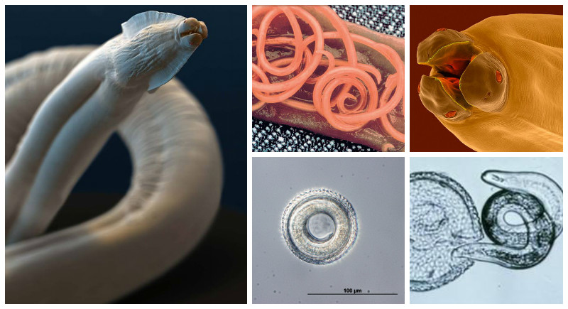
Parasites are frequent “guests” in the body of dogs, even domestic ones. In general, when pronouncing this word, many breeders may have two associations: fleas and … helminths. The last of your pet can catch, even without leaving home. Millions of years of evolution have not passed without a trace: worms have such fertility that you can easily bring a hundred or two of their eggs on their shoes. In the external environment, they are always there. The most common are round worms in dogs. They are ascarids, they are nematodes.
Contents
General information
In general, worms of this type are extremely “versatile.” They are found in the body of almost all mammals, including whales, as well as reptiles, fish, birds and amphibians. Fortunately, the most part is very specific, worms can parasitize only in the body of certain types of warm-blooded. There are, of course, “universals”, but they are few.

More than 90% of adult roundworms live in the lumen of the small intestine, but there are also species localized in the lungs, heart, blood vessels and even stomach. All parasitic nematodes are extremely prolific, and one sick pet can throw out millions of invasive eggs per day. However, these are only general characteristics of worms: many of them have extremely specific and rather complex life cycles. Therefore, the owner of the dog needs to know not only the signs of infection, but also the ways of infection, because otherwise you can hardly prescribe a full and effective therapy, and to prevent infection will be even more difficult.
In this article, it is impossible to cover even a tenth of all varieties of parasitic roundworms, and therefore consider toxocar. Their life cycle is similar to that of other parasites, and the prevalence is so high that some veterinarians say, at least, about 67% of all dogs in our country (the situation is approximately the same in the world).
Characteristics of the life cycle
There are three types of round worms, which are real “champions” in terms of prevalence in the environment.
Toxascaris leonina
Perhaps the most “unprincipled” worm, the life cycle of which is ridiculously simple. The dog only needs to swallow the invasive eggs of the parasite, after which larvae emerge from them, beginning to live and develop in the intestine of the dog. Worms are dioecious. Females that have reached puberty, actively mate and lay several hundred eggs each day (according to research results – up to 200 thousand). The latter must mature in the external environment for at least three to six weeks. But not everything is so simple.
Modern researchers believe that toxocar can not do without an intermediate host. In the role of such a small rodents most often act. Larvae hatched from eggs in their intestines, with blood flow, enter the muscle tissue of rats and mice.
Toxocaga canis
Initially “canine” worms, the development cycle is much more complicated. Moreover, these parasites have several transmission paths at once, which makes the Toxagaga canis more common in the external environment. In veterinary practice, they also have to face quite often. The pet can catch the following ways: swallowing eggs, eating an intermediate host. It is not excluded intrauterine infection and transmission of the pathogen through the mother’s milk. Let’s consider all possible ways of transfer hardly more in detail.
Swallowing of eggs. After the dog eats eggs, larvae hatch from the intestines of the animal. The latter gnaw through the walls of the digestive tract, getting into the general bloodstream. There they migrate to the lungs, but as a result they can end up in any organ and in all tissues of the canine organism. The best outcome is when the larva, encased in an unsuitable environment, is encapsulated. It’s worse when a parasite starts to grow and develop. In such situations, the liver, for example, can resemble a sea urchin, since the surface of the organ becomes literally “stuck” with worms. It is interesting that such complications are more typical for older dogs. Most likely, the body of worms is “calculated” for existence in the lungs and the gastrointestinal tract. The immune system of mature dogs (like a puppy, by the way), most likely, simply destroys the larvae that get into the liver, kidneys, other organs. In old dogs, however, the protective mechanisms do not work well.
In the lungs, the larvae remain for up to two weeks. At this time they develop and grow. At the end of this period, the body of the parasites begin to release irritating substances, causing the infected animal to develop a violent cough. Together with secreted phlegm they cough up into the mouth and swallow. Once in the digestive tract for the second time, parasites remain there forever. From the moment of infection until the moment of allocation of the first eggs with feces, it takes about a month. Note that Toxagaga canis eggs should mature in the environment for about 14 days. If the animal swallows them in an immature form, there will be no infection.
Ways of infection
- Intrauterine infection route. As we already mentioned above, the larvae trapped in the body can be encapsulated in various organs and tissues. And the organs of the reproductive system are no exception here. Larva “wake up” against the background of changes in the hormonal background, pierce the wall of the uterus and the placenta, and then find themselves in the body of an unborn puppy. This process is called intrauterine infection. Note that the larvae immediately infect the lungs of the animal. When a puppy is born, he will cough from the first days of life, coughing up and swallowing larvae, which will later develop rapidly in his digestive tract. The worst thing is that it’s not possible to treat such a young pet: you have to wait until the puppy’s weight reaches a certain minimum.
- Transmission through mother’s milk. As in the previous case, worms may well be not only in the uterus, but also in the mammary glands. When they begin to produce milk, the larvae enter the milk ducts and “go straight” directly into the digestive system of the puppies. It is interesting that during “milk” transmission, migration to lung tissue does not always take place. It is possible that in the tissues of the milk alveoli they are already ripening to the required stage. Before the mature stage, worms grow on the fourth week of the puppy’s life, after which many eggs of parasites appear in the faeces of the animal. Even if before that time there were still “pure” animals in the litter, soon after the appearance of eggs all the puppies are infected without exception.
- T. Cati. It is believed that parasites of this species in the canine organism survive physically incapable due to serious differences in the biochemistry of cats and dogs. But! There are often cases when in the stool of dogs the eggs of Tohosaga cati are found. Why this can happen? You probably will not like the answer. Dogs, as all experienced breeders probably know, often walk around the stool of other animals during walks. They do this for the sake of replenishing the “livestock” of the intestinal microflora. If the dog eats the feces of a cat or other animal infected with this type of parasite, then in its feces eggs of this species of parasitic worms may appear. Of course, some part of the larvae can hatch in the canine organism, but their development will be impossible.
Symptoms and diagnostic
As a rule, round parasitic worms feed not only with semi-digested food masses (chyme), but also with mucous membranes and blood. All this often leads to the development of severe digestive disorders. Again, with mild infection, symptoms may not appear. In other cases, the pet quickly develops marked depletion, their hair and skin become dry. In particularly severe cases, anemia appears, manifested in the form of blanching of all visible mucous membranes. Diarrhea, alternating with diarrhea, is possible.
In young animals and in severe helminthic invasion, worms can cause complete intestinal obstruction, which usually leads to the death of the pet (the body walls do not withstand such pressure and simply burst). In addition, during the migration of larvae from the lungs back into the digestive tract, pets begin to cough heavily. Finally, in the youngest and oldest animals, larvae on the “pulmonary” stage of development often cause pneumonia or, at best, severe bronchitis.
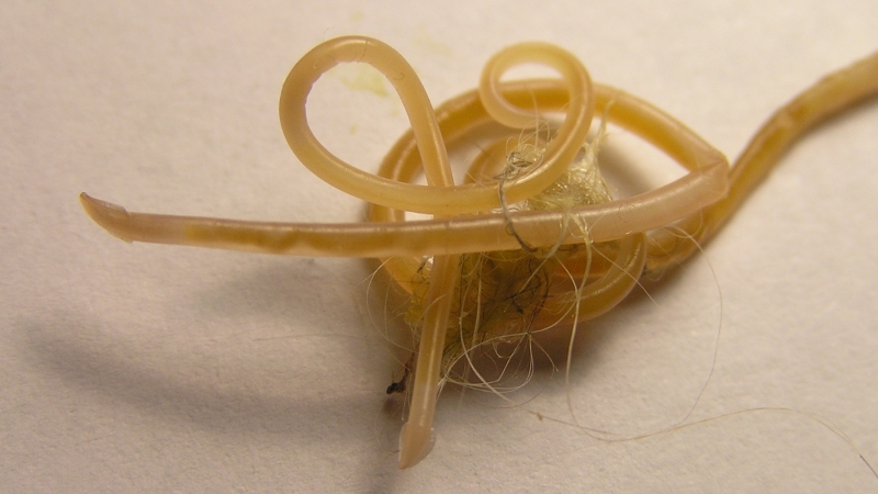
There are indications that in some animals, adult toxocar can grow up to 20 cm (although usually their size does not exceed 14 cm). If the intensity of helminthic invasion is particularly high, sometimes they can be found in fecal or vomit masses. If you do not go into details, then outwardly their clusters are like a tangle of spaghetti. But! Once again, we emphasize that this is possible only in cases of very strong helminthic invasion. As a rule, worms move very actively in the lumen of the intestine, their exit outward is almost impossible. The dead individuals are completely digested into a mucus mass, which is impossible to distinguish in feces.
So in practice, the diagnosis of “toxocarosis” (or other ascariasis) is made solely on the basis of data obtained after the analysis of stool. Typically, many species of parasitic worms are characterized by characteristic eggs that are clearly distinguishable by microscopic examination of faecal samples. However, sometimes the species is not particularly important: the veterinarian is more important to know the specific type of pathogen (round helminths in dogs suggest a different therapy if compared with cestodias).
Treatment
A full-fledged treatment does not mean merely a single administration of the drug. The fact is that in the body of the dog can always remain “sleeping” larvae. It is for this reason that in 10-14 days the therapeutic course must necessarily be repeated. In general, in the ideal case, the animal should be treated until three negative results of stool examination are obtained (in a row, of course). In such a situation, you can accurately guarantee that there are no parasites in the body of the dog. In addition, if in your area an unfavorable situation with ascaridosis, the dog’s feces should be examined at least once a month.


